Back Of Neck Anatomy / Many in the neck help to stabilize or move the head.. From the sides and the back of the neck, the splenius capitis inserts onto the head region, and the splenius cervicis extends onto the cervical region. The spine runs from the base of your skull down the length of your back, going all the way down to your pelvis. 1193).—various bony surfaces and prominences on the skull can be easily identified by palpation. Clinically, surface anatomy is used to split the neck into anterior and posterior triangles which provide clues as to the location of specific structures. The anterior jugular vein (v.
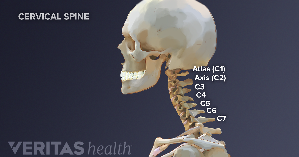
Posterior triangle of the neck boundari… pretracheal fascia b. Cervical spine anatomy video the cervical spine has 7. Neck muscles help support the cervical spine and contribute to movements of the head, neck, upper back, and shoulders. This mri neck axial cross sectional anatomy tool is absolutely free to use. The head rests on the top part of the vertebral column, with the skull joining at c1.
Cervical Spine Anatomy from embed.widencdn.net Jugularis anterior) begins near the. Want to learn more about it? Choose from 500 different sets of flashcards about neck anatomy back neck upper on quizlet. This article describes the anatomy of the head and neck of the human body, including the brain, bones, muscles, blood vessels, nerves, glands, nose, mouth, teeth, tongue, and throat. It runs from the neck to the upper back. Click now to study the muscles, glands and organs of the neck at kenhub! They control the scapulae (shoulder blades), which play a role in shrugging, neck movement, head. Dummies helps everyone be more knowledgeable and confident in applying what they know.
Choose from 500 different sets of flashcards about neck anatomy back neck upper on quizlet.
Head and neck anatomy is important when considering pathology affecting the same area. From the sides and the back of the neck, the splenius capitis inserts onto the head region, and the splenius cervicis extends onto the cervical region. It consists of seven vertebrae. Dummies helps everyone be more knowledgeable and confident in applying what they know. Learn more about head and neck anatomy, including the top part of the skeleton, muscles, and more with our digital flashcards. Dummies has always stood for taking on complex concepts and making them easy to understand. This article concerning the anatomy of the head and neck area gives you a clear structure at hand to see anatomy and function of the regions of the lower face. Posterior triangle of the neck boundari… pretracheal fascia b. Understanding the anatomy of your cervical spine and the vital nerves it contains should motivate you to adopt behaviors that help prevent neck injury and. Head and neck anatomy focuses on the structures of the head and neck of the human body, including the brain, bones, muscles, blood vessels, nerves in a newborn, the junction of the paritial bones with the frontal and occipital bones, form the anterior (front) and posterior (back) fontanelle, or soft spots. It provides images in the axial and coronal planes so that the user can study and learn anatomy. This article describes the anatomy of the head and neck of the human body, including the brain, bones, muscles, blood vessels, nerves, glands, nose, mouth, teeth, tongue, and throat. The cervical spine protects the nerves connecting to the brain, allowing the head to move freely while supporting its weight.
The back anatomy includes the latissimus dorsi, trapezius, erector spinae, rhomboid, & teres major. Posterior triangle of the neck boundari… pretracheal fascia b. « back show on map ». This entry was posted in anatomy by admin. Neck muscles help support the cervical spine and contribute to movements of the head, neck, upper back, and shoulders.
Cervical Dysfunction And Pain In The Head And Neck Causes And Osteopathic Options from www.practicalpainmanagement.com It runs from the neck to the upper back. The levator scapulae muscle is attached at the top four cervical vertebrae (c1 to c4) and runs down the side of the neck to attach at the top of the shoulder blade (scapula). Use the mouse scroll wheel to move the images up and down alternatively use the tiny arrows (>>) on both side of the image to move the images. Clinically, surface anatomy is used to split the neck into anterior and posterior triangles which provide clues as to the location of specific structures. Jugularis anterior) begins near the. Whether it's to pass that big test, qualify for that big promotion or even master that cooking technique; From the sides and the back of the neck, the splenius capitis inserts onto the head region, and the splenius cervicis extends onto the cervical region. Despite being a relatively small region, it contains a range of important anatomical features.
Click now to study the muscles, glands and organs of the neck at kenhub!
« back show on map ». The neck is the area between the skull base and the clavicles. Muscles of the posterior neck and the back. This article describes the anatomy of the head and neck of the human body, including the brain, bones, muscles, blood vessels, nerves, glands, nose, mouth, teeth, tongue, and throat. It runs from the neck to the upper back. Throughout the early years ce, and even a little back before. 1193).—various bony surfaces and prominences on the skull can be easily identified by palpation. Dummies helps everyone be more knowledgeable and confident in applying what they know. The head rests on the top part of the vertebral column, with the skull joining at c1. The neck begins at the base of the skull and connects to the thoracic spine (the upper back). Neck muscles help support the cervical spine and contribute to movements of the head, neck, upper back, and shoulders. Use the mouse scroll wheel to move the images up and down alternatively use the tiny arrows (>>) on both side of the image to move the images. The cervical spine protects the nerves connecting to the brain, allowing the head to move freely while supporting its weight.
It runs down the back part of the neck, and opens into the external jugular vein just below the middle of its course. Surface anatomy of the head and neck. The splenius muscles originate at the midline and run laterally and superiorly to their insertions. 12 photos of the anatomy of the back of the neck. Jugularis anterior) begins near the.
Cervical Spine Anatomy Neck from www.spineuniverse.com Learn everything about the neck anatomy with this topic page. The neck begins at the base of the skull and connects to the thoracic spine (the upper back). Neck muscles help support the cervical spine and contribute to movements of the head, neck, upper back, and shoulders. Learn about these muscles, their locations & functional the traps are quite a complex set of muscles. 1193).—various bony surfaces and prominences on the skull can be easily identified by palpation. The levator scapulae muscle is attached at the top four cervical vertebrae (c1 to c4) and runs down the side of the neck to attach at the top of the shoulder blade (scapula). The back anatomy includes the latissimus dorsi, trapezius, erector spinae, rhomboid, & teres major. Cervical spine anatomy video the cervical spine has 7.
It consists of seven vertebrae.
Anatomists tend to classify the body into during muscle traction, the cheeks are pulled together, which makes food move back and forth. This atlas is a comprehensive and affordable learning tool for residents and medical students and especially for radiologists and surgeons. Some important structures contained in or passing through the neck include the seven cervical vertebrae and enclosed spinal cord, the jugular veins and carotid arteries, part of the esophagus, the larynx. Head and neck anatomy is important when considering pathology affecting the same area. The head rests on the top part of the vertebral column, with the skull joining at c1. It consists of seven vertebrae. « back show on map ». It runs down the back part of the neck, and opens into the external jugular vein just below the middle of its course. The neck is the area between the skull base and the clavicles. Dummies has always stood for taking on complex concepts and making them easy to understand. From the sides and the back of the neck, the splenius capitis inserts onto the head region, and the splenius cervicis extends onto the cervical region. Surface anatomy of the head and neck. The levator scapulae muscle is attached at the top four cervical vertebrae (c1 to c4) and runs down the side of the neck to attach at the top of the shoulder blade (scapula).


0 Comments